Technology

Protonated Bioadhesive Technology (PBT™)
Bioactive Microfiber Gelling Technology (BMG™)

Axio Biosolutions employs the proprietary Protonated Bioadhesive Polymer technology (PBT) to make its biopolymer-based hemostats, drug delivery systems and scaffolds with tailorable bioadhesive properties. These preparations are unique in a sense that they adhere to the tissues when needed and easily detach when their job is done.

The essence of PBT technology lies in maximizing the bioadhesive properties of native chitosan without chemical modifications in its backbone. Structurally, chitosan is a natural polysaccharide composed of monomers of glucosamine and N-acetyl glucosamine. Chitosan gets its cationic (+ve) charge through the protonation of primary amines ( NH 2 ) groups present in its glucosamine subunits. However, this protonation is pH-dependent, at a pH above 6.5 chitosan undergoes neutralization and loses the positive charge at the physiological pH of 7.4. Researchers have developed numerous chemical derivatives of chitosan to improve its cationic properties at physiological conditions. However, such chemical modifications often lead to other drawbacks such as inconsistency, increased processing costs, risk of toxicity, and reduction in chitosan’s molecular weight. Hence, we formulated chitosan with the (PBT), which only relies on the physical properties and microscopic morphology of this natural material to maximize its cationic charge.

Axiostat is our first commercial product based on the PBT. Its uniform microscopic porous structure provides a unique molecular chemistry of chitosan within the matrix, which helps in retaining its positive charge for prolonged duration and even under physiological conditions. The highly porous structure of Axiostat helps by improving the tissue-biomaterial interaction both at macro and micro scales.

Firstly, when Axiostat is pressed against the wounded tissue it creates vacuous space within its pores which leads to mechanical interlocking with the tissue surface and provides instant bioadhesion. Secondly, the highly porous structure of Axiostat promotes the diffusion of surface-bound anionic molecules into the cationic pores of Axiostat which results in the diffusive adhesion, a mechanism in which surface bound molecules from tissues diffuse into the pores of Axiostat to form strong bioadhesion. Through this unique mechanism, Axiostat is able to maintain its positive charge and provide strong mucoadhesion even under physiological conditions.

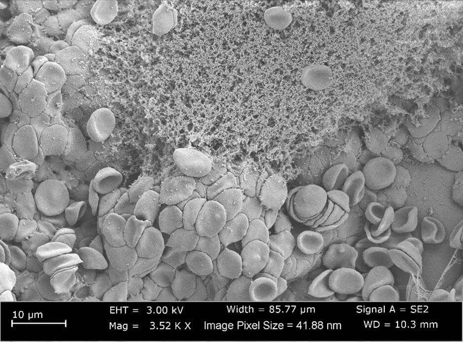
Another advantage of PBT is the faster hemostasis due to electrostatic interaction between positively charged biopolymer and the negatively charged blood cells. Through its positive charge, the PBT biopolymer attracts red blood cells (RBC) and platelets and entraps them into its porous structure, which leads to the activation of the platelets and results in formation of a strong blood clot. The combined effect of bio-adhesion and strong blood clot is instant hemostasis.
This method of bleeding control is robust enough that it works irrespective of the natural clotting factors, hence PBT is successful even in patients taking the blood thinning medications. Lastly the enhanced cationic charge provided by the PBT biopolymer also provides strong anti-microbial properties to the material and restricts entry of external bacteria into the wound site.

Chitosan promotes Haemostasis

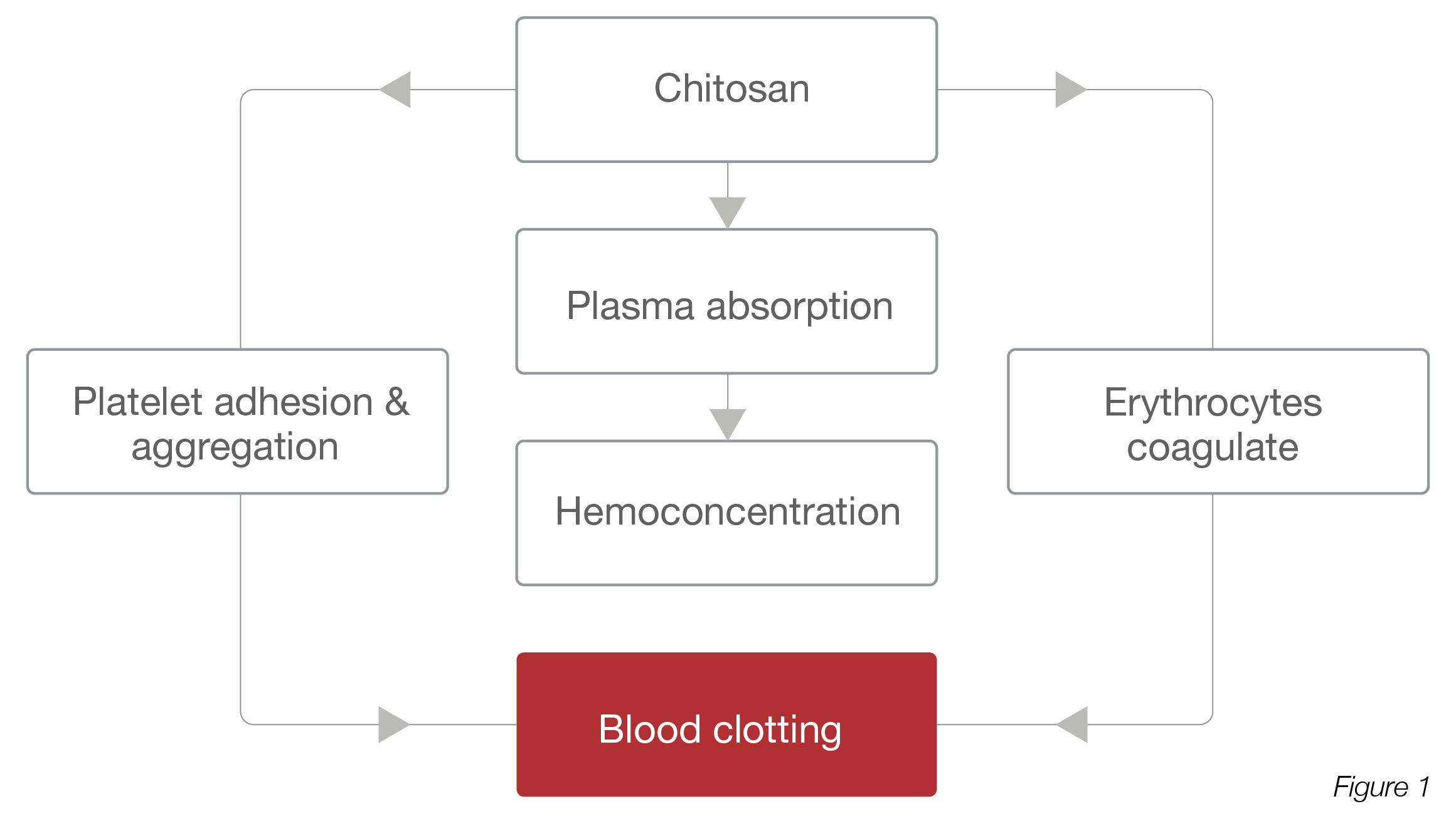
Mechanism of Haemostatic Action:

  1. Chitosan absorbs blood plasma that leads concentration of erythrocytes and platelet in the injured place.
  2. Chitosan causes the adhesion, aggregation, and activation of platelets.
  3. Chitosan promotes erythrocyte coagulation and activation due to charge-based interaction.
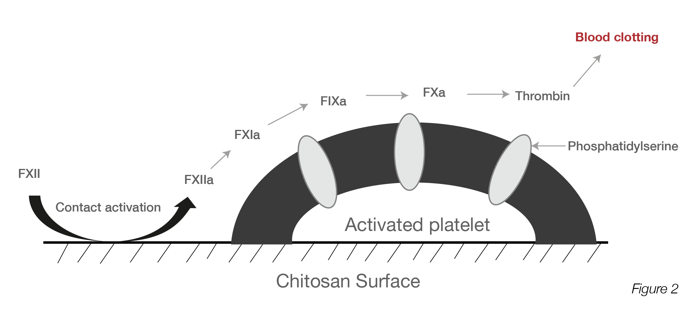
  4. Chitosan also induces blood coagulation through contact system activation
    with blood coagulation factors FXI and FXII.

Chitosan induces Platelet Aggregation, Activation and Adhesion

Mechanism of platelet activation:

  1. Platelet gets activated when chitosan binds to the glycoprotein 2b/3a receptor located on the platelet surface
  2. Chitosan promotes the platelet aggregation by stimulating the influx of extracellular Ca++ ions that cause accelerating of actin cytoskeleton activation of adhered platelets and platelet shape get changed
  3. Chitosan enhances the platelet adhesion in the presence of adsorbed plasma and extracellular matrix proteins
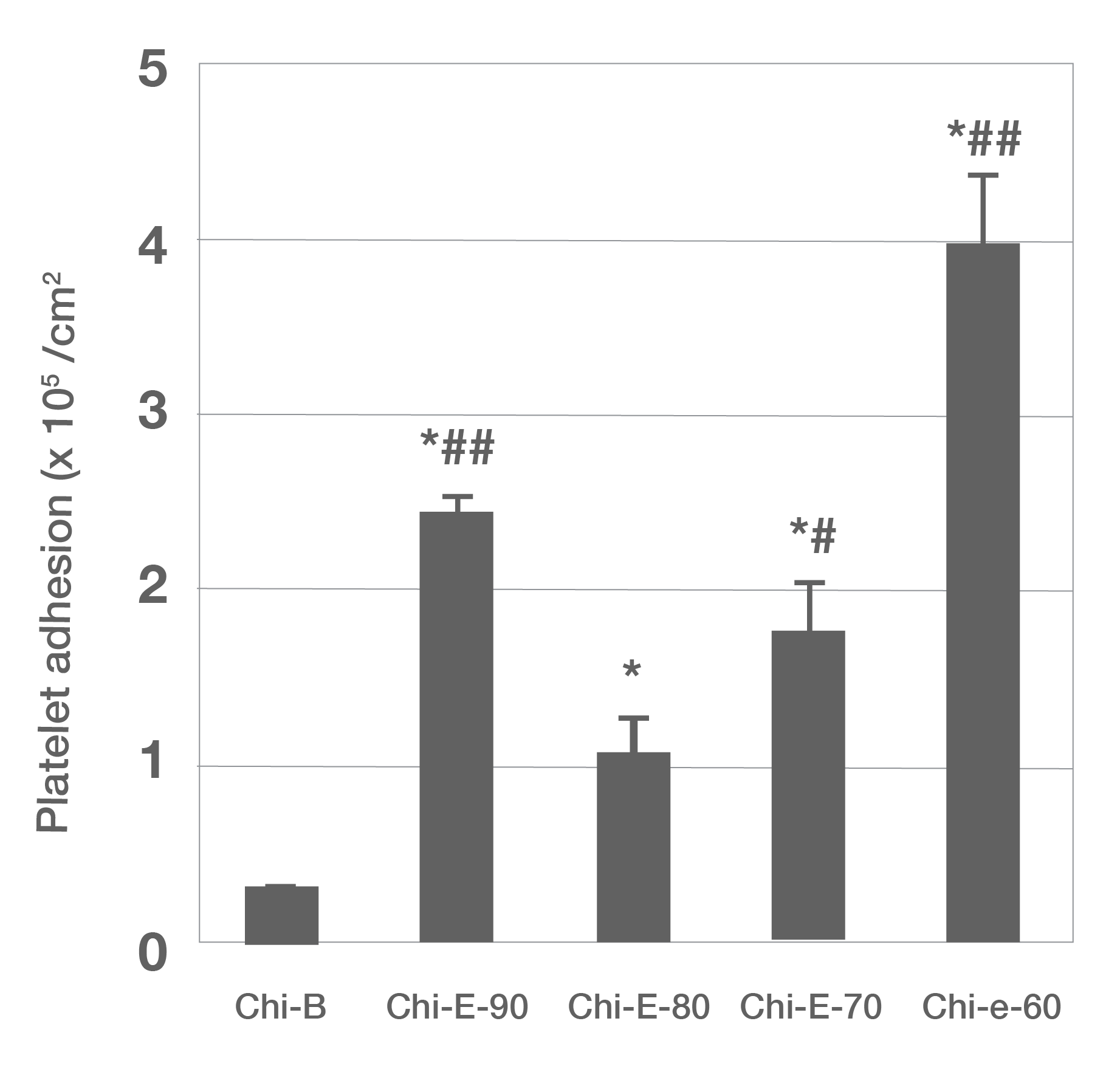
Figure 1: SEM images of platelets adhered on the chitosan dressing: a) Chi-B, (b) Chi-E-90, (c) Chi-E-80, (d) Chi-E-70, and (e) Chi-E-60. m Quantification of platelet adhesion on the chitosan films. Notes: n ¼ 4, *p < 0.001 relative to the Chi-B, # p < 0.01 relative to the Chi-E-80, # # p < 0.001 relative to the Chi-E-80 (Reproduced from Reference #2- He et al., 2013)
Chitosan causes Erythrocyte Coagulation

Mechanism of erythrocyte coagulation:

  1. Cationic chitosan can easily bind to the negatively charged neuraminic acid-containing surface of RBC due to electrostatic interaction
  2. Erythrocytes lose their biconcave shape after the interaction with chitosan that leads to agglutination of RBC
  3. Erythrocytes are bound together by chitosan polymer chains and re-polymerize into a strong lattice that capture cells creating a stable mechanical seal
Figure 1: SEM images of erythrocyte coagulation on chitosan (A), CS 6c (BJ, CS 12c (C), and CS 1 Bc (D) (Reproduced from Reference #2- Chen et al.,2017)

Evaluation of Chitosan-based Dressings in a Swine Model of Artery-Injury-Related Shock

Pre-Clinical Studies – Swine Model

Abstract:

Uncontrolled haemorrhage shock is the highest treatment priority for military trauma surgeons. Injuries to the torso area remain the greatest treatment challenge, since external dressings and compression cannot be used here. Bleeding control strategies may thus offer more effective haemostatic management in these cases. Chitosan, a linear polysaccharide derived from chitin, has been considered as an ideal material for bleeding arrest.
This study evaluated the potential of chitosan-based dressings relative to commercial gauze to minimize femoral artery haemorrhage in a swine model. Stable Haemostasis was achieved in animals treated with chitosan fibre (CF) or chitosan sponge (CS), resulting in stabilization of mean arterial pressure and a substantially higher survival rate (100% vs. 0% for gauze). Pigs receiving treatment with CF or CS dressings achieved Haemostasis within 3.25±1.26 or 2.67±0.58min, respectively, significantly more rapidly than with commercial gauze (> 1 00min). Moreover, the survival of animals treated with chitosan-based dressings was dramati­cally prolonged (> 180min) relative to controls (60.92±0.69min). In summary, chitosan-based dressings may be suitable first-line treatments for uncontrolled haemorrhage on the battlefield and require further investigation into their use as alternatives to traditional dressings in prehospital emergency care.

Figure 1: The haemostatic outcomes of CF and CS dressings compared to commercial gauze application in a swine model of arterial haemorrhage. Median time to Haemostasis. Error bars represent the 95% confidence interval of the median. The chitosan-based dressing promoted Haemostasis significantly greater than commercial gauze (Reproduced from Reference #1-Wang et al., 2019)

Outcome CF(N=4) CS(N=3) Gauze(N=3)) Overall p
Total time until bleeding stopped(min) 3.25+1.26 2.67+0.58 >20 NS
Total resucitation fluid (mL/kg) 30.55 31.58 188.32 <0.001
Survival rate(%) 100% 100% 0% NS
Survival rate(min.) >180 >180 60.92+0.69 NS

Table 1: Outcomes of treating a groin arterial haemorrhage. Data expressed as mean±SD and analyzed by one way ANOVA. NS=p>0. 1. ANOVA, analysis of variance; NS, not significant.

Blood Clot Test

Axiostat Vs. Other Haemostatic Gauze

REFERENCES

  1. Maksym P\/, Vitalii S. Chitosan as a hemostatic agent: current state. Eur J Med Ser B. 2015;2(1 ):24-33.
  2. Hu Z, Zhang DY, Lu ST, LJ PW, LJ SD. Chitosan-based composite materials for prospective hemostatic applications. Marine drugs. 2018 ;16(8):273.
  3. He Q, Gong K, Ao Q, Ma T, Yan Y, Gong Y, Zhang X. Positive charge of chitosan retards blood coagulation on chitosan films. Journal of biomaterials applications. 2013;27(8):1032-45
  4. Zhou X, Zhang X, Zhou J, Li L. An investigation of chitosan and its derivatives on red blood cell agglutination.
    RSC Advances. 2017; 7(20): 1224 7 -54.
  5. Chen Z, Yao X, Liu L, Guan J, Liu M, Li Z, Yang J, Huang S, Wu J, Tian F, Jing M. Blood coagulation evaluation of N-alkylated chitosan. Carbohydrate polymers. 2017; 173:259-68
  6. Arand AG, Sawaya R. lntraoperative chemical HAEMOSTASIS in neurosurgery. Neurosurgery. 1986 Feb 1; 18(2):223-33.
  7. Wang YH, Liu CC, Cherng JH, Fan GY, Wang YW, Chang SJ, Hong ZJ, Lin YC, Hsu SD. evaluation of chitosan-based
    Dressings in a Swine Model of Artery-injury-Related Shock. Scientific reports. 2019 ;9(1 ): 1 -7.

Speak to the team

Our office is open Monday - Friday 8.30am - 5.00pm

Feel free to get in touch direct or leave us a message on the contact form and the team will get back to you

Phone

+44 (0) 1934 53 34 53

Address

65 Bath Road, Stroud, Gloucestershire, GL5 3LA